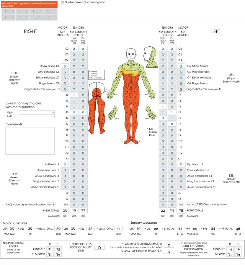
Printable Spinal Cord Assessment and Dermatone – Spinal cord assessment is a crucial part of any neurologic evaluation. It allows doctors to determine the extent of spinal cord injuries and diagnose underlying conditions that affect the nervous system. The assessment involves examining the patient’s anatomy, assessing motor and sensory functions, and testing reflexes.
One way to assess spinal cord function is through a printable spinal cord assessment form. This form enables physicians to keep track of their patient’s progress over time and helps them make informed decisions about treatment plans. Additionally, dermatome maps can be used in conjunction with spinal cord assessments as they indicate which areas on the skin corresponding with specific nerves in the body.
Overall, performing regular spinal cord assessments is essential for ensuring optimal health outcomes for patients with neurological conditions or injuries affecting this critical part of their body. By using standardized tools like printable forms and dermatome maps, medical professionals can stay organized, accurate, and efficient in their diagnoses and treatments.
How Many Spinal Nerves Are In Your Dermatome?
Dermatomes are areas of the skin that are innervated by a single spinal nerve. The human body has 31 pairs of spinal nerves, and each pair serves a specific dermatome. This means that there are 62 dermatomes in the human body.
Each spinal nerve is responsible for a specific region of the body, and these regions can overlap. For example, the C5 dermatome covers part of the shoulder and upper arm, while the C6 dermatome covers part of the forearm and thumb.
Understanding dermatomes is important in diagnosing certain medical conditions that affect nerve function. By mapping out which areas of the skin are affected, doctors can determine which spinal nerves may be damaged or compressed.
The printable spinal cord assessment and dermatone chart can be helpful tools in identifying potential nerve damage or compression. By marking off areas on the chart where a patient experiences pain or numbness, doctors can narrow down which spinal nerves may be involved and provide appropriate treatment.
What Are The Dermatomes Of The Sacral Spine?
Dermatomes are areas of the skin that are innervated by specific spinal nerves. The sacral spine, which is located at the bottom of the vertebral column, has five dermatomes: S1 to S5. Each dermatome corresponds to a specific area on the skin and can be used to assess nerve function in patients with suspected spinal cord injury or disease.
Assessing dermatomal sensation is an important aspect of neurological examination. A printable spinal cord assessment and dermatome chart can be useful for healthcare professionals to document their findings during physical exams. In addition, understanding the distribution of sacral dermatomes can also aid in identifying potential sources of pain or sensory deficits in patients with lower back pain or leg numbness.
Overall, knowing the location and function of sacral dermatomes is essential for healthcare professionals involved in spinal cord assessment and diagnosis. Utilizing a printable chart can help streamline documentation and ensure accurate communication between healthcare team members regarding patient assessments.
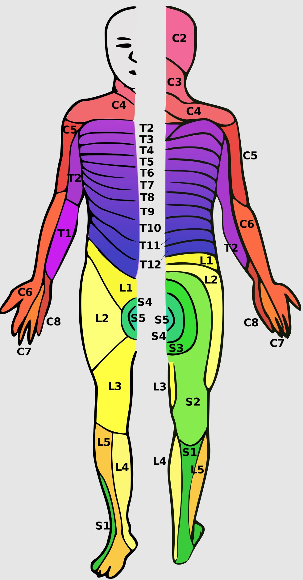
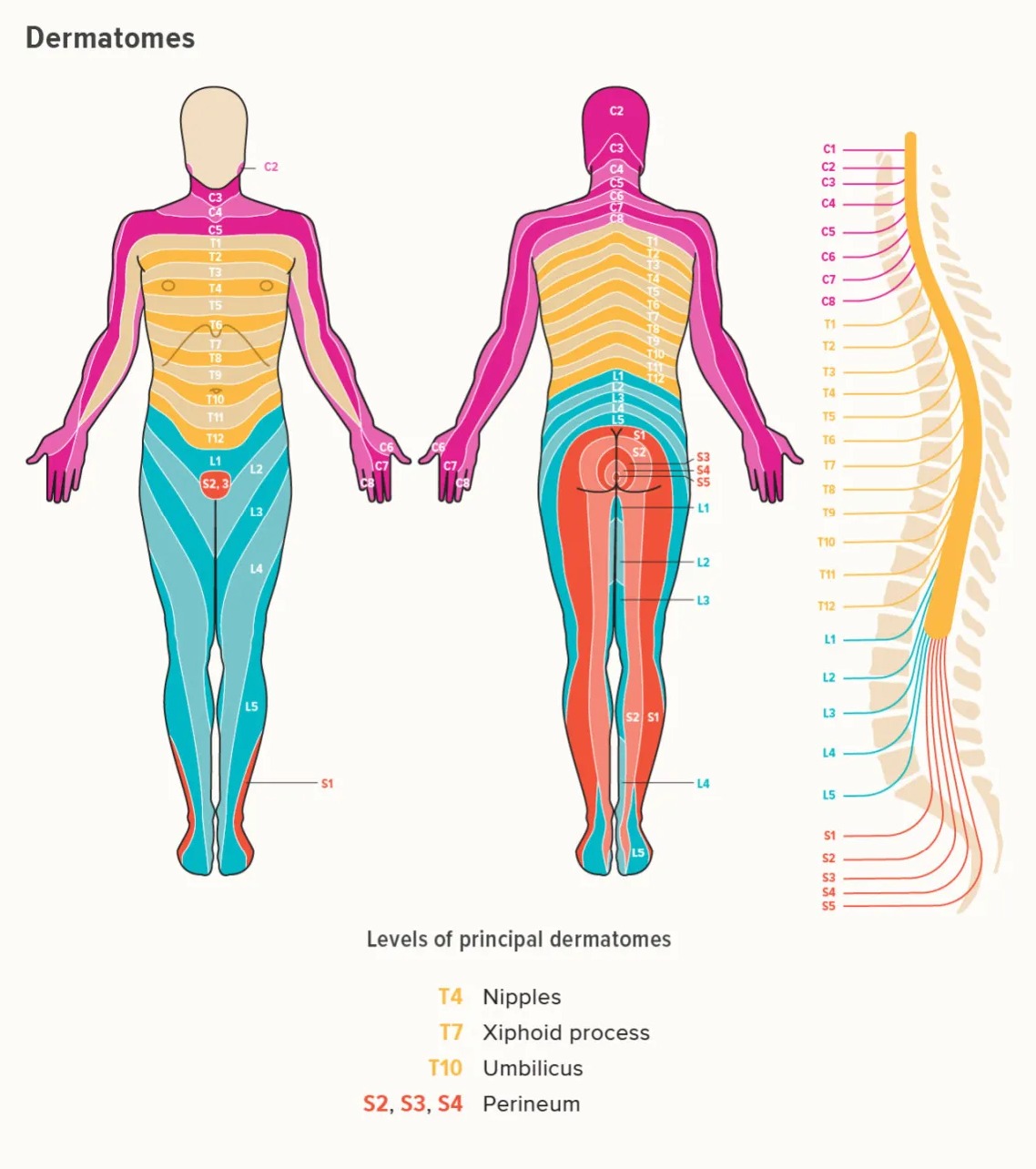
Printable Spinal Cord Assessment and Dermatone
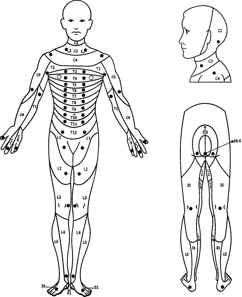
A printable spinal cord assessment and dermatome chart is a useful tool for healthcare professionals to assess and diagnose spinal cord injuries. The chart shows the different levels of the spinal cord and their corresponding sensory and motor functions. It can be used to identify which part of the spine is injured and what type of symptoms the patient may experience.
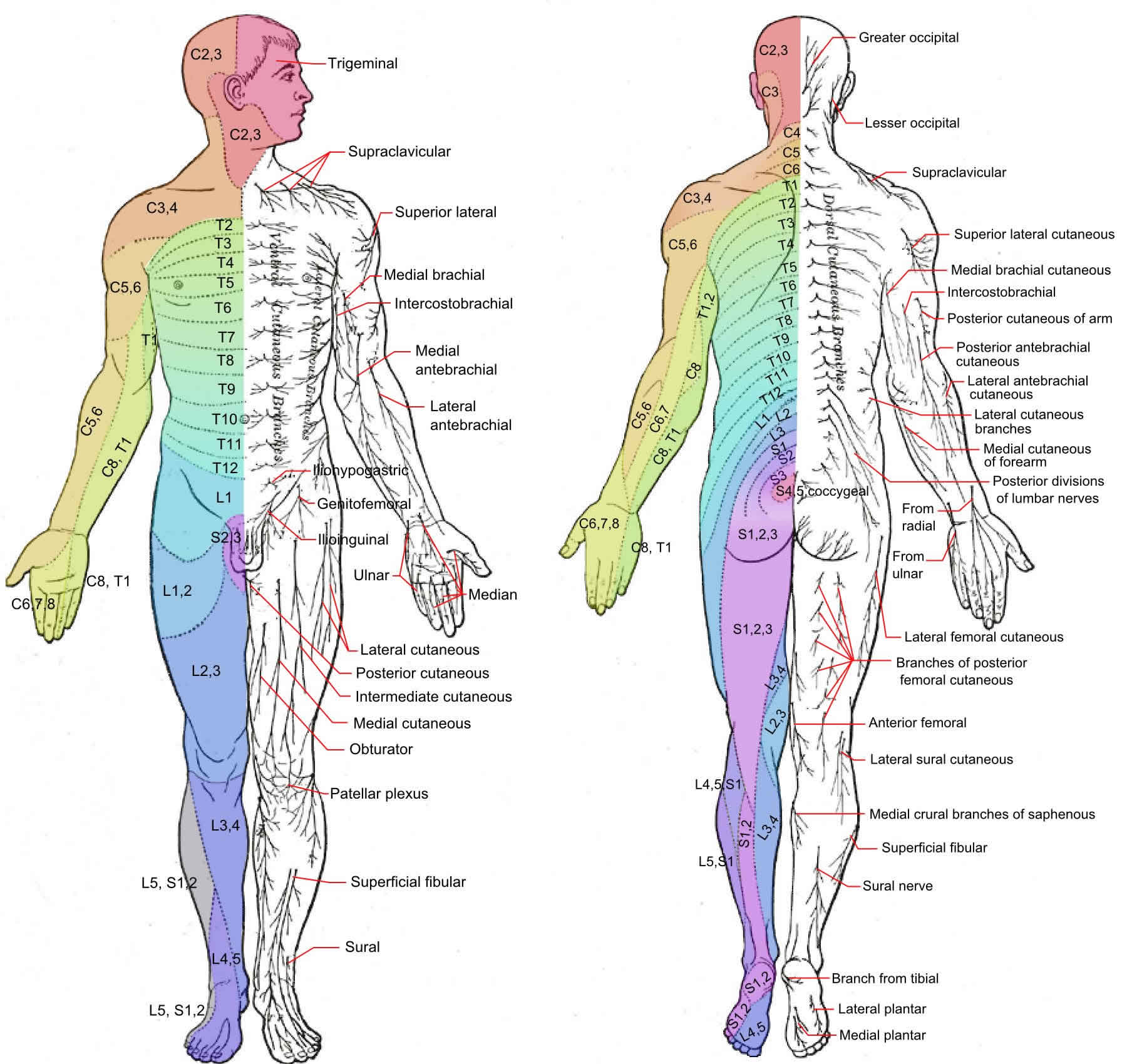
The dermatome chart on the same page shows which areas of skin are supplied by each spinal nerve. This information is important in diagnosing neurological disorders that affect sensation or pain perception in specific parts of the body.
Having a printable version of these charts can be handy for healthcare professionals who need quick access to this information during patient assessments. It also serves as a helpful visual aid when explaining diagnoses to patients, allowing them to understand where their symptoms are coming from and what they can expect during treatment.


